1. Introduction and Background
Renal cell carcinoma (RCC) is frequently detected incidentally on imaging tests performed for other reasons, and its incidence continues to increase, particularly in the elderly (1). Although surgical resection of the tumor remains the treatment of choice for clinically localized disease in the majority of otherwise healthy of patients, it may not be the ideal or optimal approach for all. Additional patient related factors such as frailty, reduced renal function, anatomical limitations, age, or even patient refusal of surgery have prompted the search for therapeutic alternatives (2). For patients with small renal masses less than 3-4 cm in diameter, active surveillance is increasingly considered an acceptable strategy, as many such patients will likely never require surgery for benign or indolent tumors (3). On the other hand, a subset of these individuals will progress during active surveillance and ultimately require local intervention. Minimally invasive strategies to avoid treatment related morbidity and to preserve renal function include focal therapies such as cryoablation (CA) and radiofrequency ablation (RFA), but these methods are best used for carefully selected small tumors in favorable locations, away from the bowel, urinary tract, and great vessels (4). Significant gaps remain for patients with larger tumors, complex anatomy, solitary kidneys, or other medical contraindications for even minimally invasive surgery.
Radiotherapy (RT) has historically had a limited role in RCC because the disease has long been considered inherently radioresistant, which led to radiotherapy being largely restricted to palliative treatment of symptomatic advanced disease. However, in recent years this landscape has changed with the introduction of stereotactic ablation radiotherapy (SABR), also called stereotactic body radiotherapy (SBRT), into clinical practice. SBRT delivers highly conformal, ablative doses with steep dose gradients delivered in a small number of fractions. Ablative radiotherapy has been made possible by advances in imaging, treatment planning, delivery equipment, and respiratory motion management (5, 6). Together, these advances mark a major transition from the historically limited role of conventional radiotherapy in RCC to modern SBRT/SABR as a noninvasive ablative strategy for selected patients unsuitable for surgery (Figure 1).
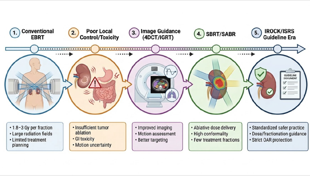
Figure 1. Historical evolution of radiotherapy for primary renal cell carcinoma. Conventional radiotherapy was limited by low biologically effective dose, poor image guidance, respiratory motion, and gastrointestinal toxicity. Modern SBRT/SABR has enabled ablative dose delivery with improved local control and acceptable toxicity.
Mechanistically, preclinical work suggests that high-dose, hypofractionated (use of higher doses of radiation per session in fewer total treatments than conventional radiation) radiotherapy may overcome the potential radioresistance of RCC, possibly by engaging pathways associated with HIF1α activation in VHL-mutated clear cell RCC and vascular/endothelial effects beyond classical DNA damage mechanisms (7). Clinical studies demonstrate that SBRT can achieve highly promising local control with an acceptable toxicity profile and preservation of renal function, allowing radiotherapy to be increasingly integrated into multidisciplinary care for selected patients, including those with more advanced local disease where sustained local control is desired (8, 9)
2. RCC Biology and Radioresistance
Historically, RCC was treated with conventional fractionated external beam radiotherapy, typically 1.8–3.0 Gy per fraction, delivered in multiple sessions, with consistently unsatisfactory results. This was reinforced by the mistaken assumption that all radiotherapy treatments are biologically equivalent and that changing the dose per fraction or delivery technique would not substantially alter tumor response. Radiotherapy with small daily fractions relies mainly on induced oxygen-dependent DNA damage and can induce hypoxia-reoxygenation cycles that increase ROS, HIF1A signaling, and various angiogenic factors such as VEGF/FGF, enhancing a radioresistant phenotype in this highly vascular tumor and thus rendering this approach ineffective. Furthermore, microvascular endothelial apoptosis appears to play a crucial role in the effective control of RCC tumors and is not successfully activated by conventional low-dose fractions, resulting in insufficient vascular destruction and tumor eradication. Finally, the use of large abdominal fields as well as attempts to scale up fractionation with older, non-axial/non-image-guided techniques resulted in significant irradiation of the small bowel, with no survival benefit and high severe toxicity, including radiation-related deaths (7).
Understanding the biology of ccRCC provides important context for how loss of VHL function leads to an imbalance between hypoxia/angiogenesis and shapes the response to radiation (Figure 2). As a substrate recognition unit of an E3 ubiquitin ligase, under normoxic conditions, pVHL promotes PHD-dependent hydroxylation of HIF-1α, leading to its ubiquitination and proteasomal degradation. When VHL function is lost in ccRCC, hydroxylation is reduced, HIFα accumulates, dimerizes with HIF-1β/ARNT, associates with hypoxia response elements, and leads to a sustained pseudohypoxic transcriptional state that also rewires metabolism towards aerobic glycolysis and glutamine-mediated reductive carboxylation.
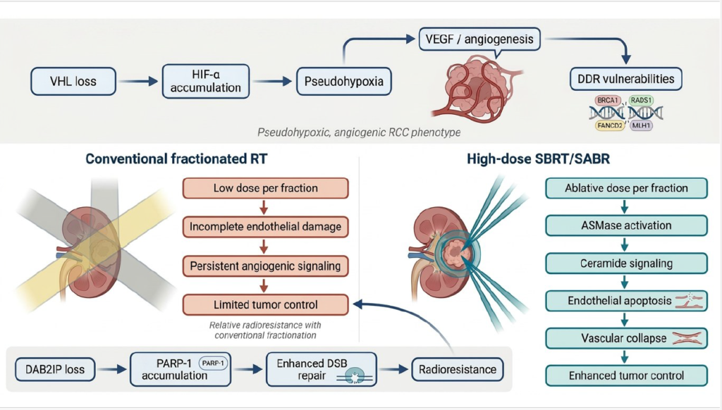
Figure 2. Proposed biological mechanisms underlying RCC radioresistance and response to ablative radiotherapy. VHL loss and HIF activation promote a pseudohypoxic and angiogenic phenotype. High-dose SBRT may overcome conventional radioresistance through endothelial apoptosis, vascular collapse, and enhanced tumor control.
Loss of oxygen-dependent proteasomal degradation of HIFα mediated by normal pVHL gene function, which is inactivated in more than 60% of ccRCC, results in a sustained, constitutive increase in HIFα, establishing a hypoxia-like transcriptional context. Genomic instability is thought to be primarily a consequence of hypoxia, driven by the suppression of DNA repair pathways through transcriptional, translational, and epigenetic mechanisms. Thus, loss of VHL may similarly create a vulnerability to the DNA damage response (DDR) by repressing repair genes. Indeed, in ccRCC cell lines, VHL–/– cells display reduced homologous recombination at BRCA1, RAD51, FANCD2, and mismatch repair at MLH1, both at the mRNA and protein levels. In particular, transient VHL silencing in VHLWT cells reproduces this phenotype, linking it directly to pVHL loss rather than cell cycle differences. VHL–/– cells are functionally more sensitive to radiation and to PARP inhibitors, and repair double-strand breaks induced by ionizing radiation less efficiently. Specifically, the pattern of VHL-inactivated tumors shows lower FANCD2, BRCA1, RAD51, and MLH1 with higher hypoxia markers, such as VEGFA. The mechanism likely involves multiple hypoxia regulators, suggesting that HIF-1 is redundant and that HIF-2 may play a greater role in DDR suppression (10).
In ccRCC, a subpopulation of radiation-resistant tumor-initiating cells (TICs) can survive fractionated therapy by enhancing checkpoint signaling and arresting the cell cycle for repair. Indeed, one study reported that in freshly isolated primary human ccRCC cells treated with clinically modeled fractionation at 3 Gy × 1–3 days, the growing fractions were particularly enriched for TIC-like cells, as demonstrated by increased formation of free-floating spheres in low-adhesion culture and by upregulation of stemness genes, such as Bmi1, Nanog, Sox2, Oct4. Indeed, cells that survived fractionated radiation also showed greater carcinogenesis and higher tumor burden in vivo in NOD/SCID mice. Analyzing the mechanism, it appears that fractionated radiotherapy increases ATM, Chk1 and Chk2 checkpoint transcripts, consistent with dependence on the ATM-Chk axis after ionizing radiation injury. This is thought to lead to significant G2/M arrest. In fact, inhibition of checkpoint kinases with AZD7762 reduced the clonal survival of irradiated survivors and abolished the G2 checkpoint, effectively removing the advantage of pause and repair (11).
DAB2IP-negative cells exhibit higher clonal survival after ionizing radiation, and re-expression of DAB2IP resensitizes them, demonstrating that loss of the DAB2IP tumor suppressor is associated with a radioresistant RCC phenotype. In fact, cells with low DAB2IP repair double-strand breaks much more rapidly, as shown by γH2AX kinetic analyses, suggesting that the resistance is due to increased repair efficiency rather than reduced damage induction. Specifically, DAB2IP interacts with PARP-1 and represses PARP-1 protein and activity through post-translational control without significant changes in key HR/NHEJ factors, making PARP-1 a key DDR-modified node. Indeed, to promote PARP-1 ubiquitination and proteasomal degradation, DAB2IP acts as a scaffold that recruits E3 ligases, including RanBP2, TRIP12, and RNF40. Therefore, loss of DAB2IP allows for PARP-1 accumulation, stronger checkpoint/repair signaling, and faster DSB repair. Therapeutic data support this reasoning as PARP-1 overexpression induces resistance, PARP-1 silencing restores radiosensitivity, and PARP inhibition sensitizes radioresistant RCC to radiation in vitro and improves tumor control with radiotherapy in xenograft models. Thus, DAB2IP and PARP-1 are potential biomarkers for patient selection to identify those most likely to benefit from combining a PARP inhibitor (PARPi) with radiotherapy (RT) (12) (Figure 2).
3. Results of Conventional Radiotherapy
Historically, traditional EBRT (external beam radiation therapy) was limited by conventional fractionation at 1.8–3 Gy/fraction and modest total doses such as 30–33 Gy preoperatively or 50 Gy postoperatively. This approach was primarily conservative rather than targeted biologically effective, resulting in ineffective eradication of RCC and perpetuating the perception of RCC radioresistance as dose escalation was limited by normal tissue tolerance. This was contributed by various technical limitations in imaging and treatment planning, such as imprecise target and organ at risk (OAR) definition, and limited beam shaping that forced large irradiation margins and irradiation of more normal tissue to ensure coverage. Additionally, respiratory movement of renal tumors led to further widening of the radiation margins and increased dose to nearby organs such as the bowel, stomach, duodenum, liver, spinal cord, and contralateral kidney, making toxicity a major obstacle to intensifying radiotherapy (5). Randomized trials and meta-analyses from the past on the use of adjuvant radiotherapy for RCC have generally not shown a clear benefit in either overall survival (OS) or local control. Indeed, it is readily apparent in the literature that gastrointestinal toxicity was a major limitation because the treatment fields exposed the upper abdominal organs. In particular, in the randomized study by Kjaer et al. using 50 Gy in 20 fractions for stage II and III RCC, gastrointestinal toxicities involving the stomach, duodenum, and liver were reported in 44% of patients, with a high mortality rate in the radiotherapy arm (13).
4. Improvements in Radiation Delivery Leading to SABR
Substantial technological advances in imaging, immobilization, treatment planning, and image guidance drove the transition from conventional radiotherapy to SBRT/SABR. Older techniques suffered from imprecise target and OAR definitions, limited beam shaping, and an inability to adequately account for the respiratory motion of renal tumors. These limitations necessitated large treatment fields and margins, which increased radiation dose to surrounding organs such as bowel, stomach, duodenum, liver, spinal cord, and contralateral kidney, making toxicity a major barrier to dose escalation (5). Modern renal SBRT/SABR therefore depends on an integrated workflow of careful patient selection, motion-informed simulation, strict organ-at-risk constraints, image-guided delivery, and structured renal function monitoring (Figure 3).
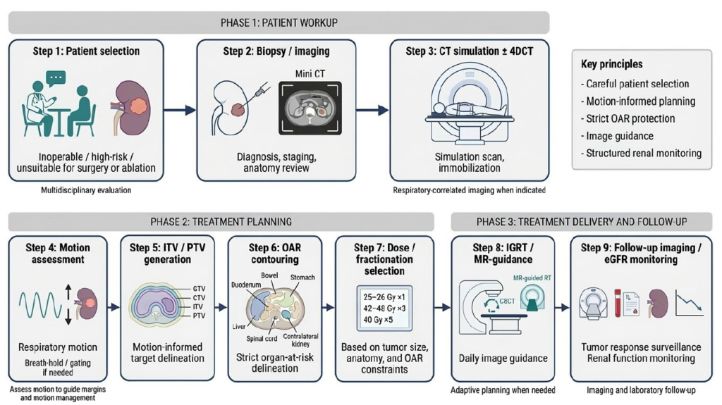
Figure 3. Practical workflow for SBRT/SABR in primary renal tumors. Safe renal SBRT requires careful patient selection, motion-informed planning, strict organ-at-risk constraints, image guidance, and structured monitoring of renal function.
The literature supports that tumor control by radiotherapy is not solely due to direct tumor cell killing, but is also largely due to apoptosis of tumor microvascular endothelial cells, with the apoptotic capacity of the patient’s endothelium determining both tumor growth and radiosensitivity. Indeed, in mouse models, endothelial apoptosis occurred within hours and preceded tumor cell apoptosis, with higher doses of about 20 Gy partially overcoming tumor resistance (14). Tumor response to radiation often depends on the vascular and endothelial microenvironment, with the dominant mechanism varying with the dose per fraction. Specifically, during conventional fractionation, hypoxia-reoxygenation cycles generate ROS that activate HIF-1 and increase VEGF and other pro-angiogenic signals, thereby protecting the endothelium and promoting radioresistance. In contrast, SBRT, due to its large fractions of 15–20 Gy, triggers a rapid, asmase- and ceramide-mediated wave of endothelial apoptosis that precedes tumor cell death. Specifically, high-dose radiation rapidly activates asmase, which converts sphingomyelin to ceramide, which in turn accumulates in endothelial cell membranes, thereby inducing apoptosis. The result of this vascular damage is microvascular collapse and reduced blood flow to the tumor, resulting in nutrient deprivation, control, and ultimately tumor cell death (15).
An additional limitation of classical irradiation of renal tumors is the movement of the kidneys due to respiration. Especially in SBRT, where the dose delivered per fraction is high, and margins are narrow, particularly in the superior-inferior (SI) direction, there is a risk of underdosing the target or overdosing the OARs. For this reason, guidelines recommend respiratory management when motion is ≥5 mm. Specifically, in a study using 4DCT (a CT scan across the breathing cycle) to quantify motion and kV-CBCT (cone-beam CT) with 6DoF couch corrections to measure residual error in patients immobilized primarily with BodyFIX (a vacuum cushion system), thirty patients showed mean inspiratory-expiratory motion of 1.5 mm (LR), 8.1 mm (SI, with some >10 mm), and 3.1 mm (AP), while a subset of fifteen patients had small residual errors that were even more common. With the above techniques, positional accuracy of 2.1 mm and rotational accuracy of 0.8° were achieved, supporting the practicality of an isotropic PTV margin of 5 mm and emphasizing the value of rotational correction and, possibly, breath holding or fencing to further reduce the treated volume and protect the adjacent OARs (16).
A retrospective study included three elderly, medically inoperable patients with stage I clear cell RCC measuring 3.6 to 5.7 cm and stage 3 and 4 CKD, with one patient having a solitary kidney, to assess feasibility, toxicity, local control, and renal function. Specifically, patients underwent tight immobilization during CT planning and PTV (Planning Target Volume) margins of 1–3 mm, abdominal compression to reduce respiratory motion, spinal tracking for any motion or the use of special implanted fiducials that detect tumor motion and adjust the beam to respiration, and received 40 Gy in 5 fractions. As a result, all tumors remained locally stable over a period of 12 to 40 months, with only acute grade 1 symptoms and no late grade 3, 4 toxicity or need for dialysis. However, renal function deteriorated over time, particularly in the patient with initial stage 4 CKD, who progressed to stage 5 at 26 months, while the two with stage 3 showed moderate deterioration (17).
Currently, adaptive MR SBRT (MRgRT) represents the next step in RCC treatment planning, because conventional CBCT (cone-beam CT)/4DCT (four-dimensional CT)-ITV (Internal Target Volume) therapeutic approaches provide only limited soft-tissue contrast and therefore may not highlight small renal displacements that are important. Thus, the MRI-guided method offers improved imaging, better real-time monitoring with beam control activated only when the target is in the correct position, often with breath holding, and continuous reoptimization of the target plan for the patient’s specific anatomy. This makes it a particularly useful therapeutic approach for patients where the possibility of irradiating a normal kidney or a normal surrounding organ is now minimized (18). A recent study supports the use of MRI-guided SBRT to address the main challenges of SABR in primary RCC. Specifically, a prospective single-center study of 36 mainly elderly patients, with a mean tumor size of 5.6 cm, investigated intra-fragmentary kidney motion and its proximity to the peri-mobile gastrointestinal organs using high-resolution soft-tissue MRI with real-time monitoring and online reoptimization as the anatomy shifted. Patients were treated with an MRI-guided radiation system with a 0.35 Tesla scanner, a method that allowed operators to observe the tumor and nearby organs during treatment, and received a total of 40 Gy over 5 treatments with simultaneous respiratory motion control. This therapeutic approach resulted in 95% local control and 91% overall survival at 1 year, with very low toxicity and a mean eGFR decline of only 6 mL/min/1.73 m², without the need for dialysis (19).
A prospective Phase I trial (NCT02264886) was undertaken because abdominal SBRT often encounters gastrointestinal OARs that shift with respiratory motion, and MR-guided, online adaptive SBRT (SMART) was hypothesized to improve target coverage and OAR preservation compared with non-adaptive SBRT. In 20 evaluable patients with oligometastatic or unresectable primary abdominal malignancies (10 hepatic, 10 extrahepatic), 50 Gy/5 fractions were administered primarily with strict isotoxicity and cine-MR gating during expiration while free-breathing. An adapted plan was superior in 83.5% of fractions, and all patients required adaptation in at least one fraction (100% of extrahepatic fractions were adapted, versus 66% of hepatic fractions). The primary reason for adaptation was to avoid violating OAR constraints, and the adapted designs prevented 100% of potential violations. Οverall cumulative GTV(Gross Tumor Volume)/PTV(Planning Target Volume) coverage was better with SMART than with the non-adaptive design, although some fractions required PTV deescalation for OAR. Clinically, there was no acute GI toxicity greater than grade 3, quality of life did not deteriorate, local stability and control were high (95% at 3 months, 89% at 6 months, only 2 local progressions in 15 months), and 1-year OS was 75% overall (91% in oligometastatic) (20) (Figure 3).
5. IROCK and the Standardization of Safer Practice
The International Consortium for Radiation Oncology for Kidney Study (IROCK) for Primary Renal Cell Carcinoma aimed at standardizing safe SBRT for primary RCC. Overall, IROCK defines the essentials for safer practice (movement-informed planning, realistic margins, strict IGRT, emphasis on GI restrictions, and structured follow-up), while recognizing major gaps that require multi-institutional and prospective trials for renal function thresholds, optimal dose–fractionation, margin strategies, and combinations with TKIs, immunotherapies, and better response assessment tools (21). IROCK is especially important because it moves the field beyond proof of concept and toward safer, more reproducible practice. At the same time, it also highlights major areas where evidence remains limited, including renal function thresholds, optimal dose-fractionation schedules, ideal margin strategies, integration with tyrosine kinase inhibitors or immunotherapy, and improved tools for assessing treatment response.
6. Modern Results of SBRT/SABR: Efficacy and Safety
SBRT/SABR has emerged as a noninvasive treatment option for selected patients with primary renal cell carcinoma, particularly those who are medically inoperable or poor surgical candidates. Its main clinical advantage is the ability to achieve durable local tumor control while preserving renal parenchyma. Response to this treatment may be slow, and assessment of treatment is generally most reliable after 6 to 12 months. Routine post-treatment biopsy is not usually required unless there is concern for true disease progression. Current Radiosurgery Society guidance supports use even in patients with a solitary kidney when renal dose constraints and follow-up are carefully managed, because dialysis is uncommon and eGFR decline is usually modest. More complex scenarios, such as tumor thrombus involving the inferior vena cava, require individualized multidisciplinary decision-making and coordination with systemic therapy (9). The major contemporary studies evaluating renal SBRT/SABR, including study design, dose/fractionation, treatment-planning or motion-management features, local control, renal-function outcomes, severe toxicity, and dialysis risk are summarized in Table 1.
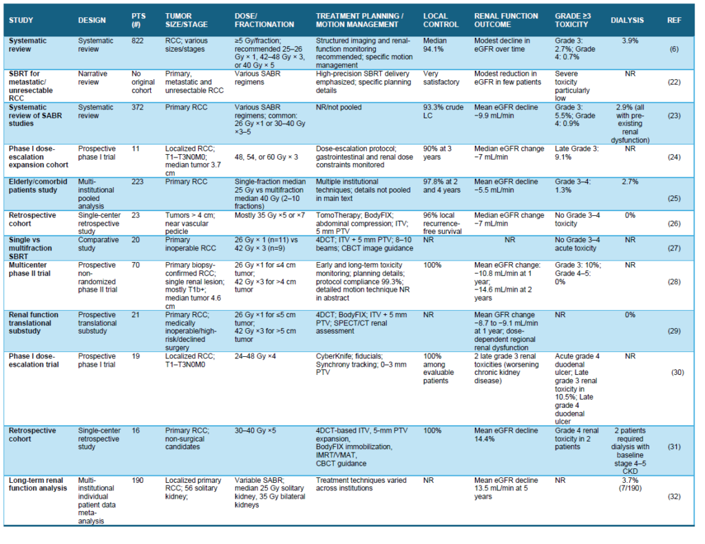
Table 1. Clinical outcomes, renal safety and treatment-planning features of renal SBRT/SABR studies
Across contemporary studies, renal SBRT/SABR demonstrates high efficacy and acceptable safety. A systematic review of 822 patients[AP1] [DS2] treated with renal SBRT at≥5 Gy per fraction reported a median local control rate of 94.1%, a 5-year progression-free survival of 80.5%, and a 5-year overall survival of 77.2%. Toxicity was limited, with dialysis required in 3.9% of patients and grade 3–4 toxicity reported in 2.7% and 0.7% of patients, respectively. The included cohort was older and medically vulnerable overall, with a mean age of 71.7 years, a median baseline estimated glomerular filtration rate of 55 mL/min, and a median maximum tumor size of 4.4 cm. Biopsy confirmation was common, with a median rate of 98% across reporting studies, and the median follow-up was 31.2 months. The authors recommended 25–26 Gy in 1 fraction for smaller tumors when organ-at-risk constraints allow, 42–48 Gy in 3 fractions for larger tumors, and 40 Gy in 5 fractions when 3-fraction constraints cannot be met, along with pretreatment biopsy and structured renal monitoring (6).
SBRT has also shown favorable activity in unresectable primary RCC, metastatic RCC, oligometastatic or oligoprogressive disease, and as a strategy to defer or extend systemic therapy, with stereotactic radiosurgery preferred for brain metastases (22). Renal function outcomes are generally favorable but remain a key safety endpoint. In the literature, primary RCC SABR is associated with low severe toxicity, with grade 3 and grade 4 events reported in 5.5% and 0.9%, respectively, and no grade 5 events. Mean baseline eGFR was 57.5 mL/min, with an average post-treatment decline of approximately −9.9 mL/min. Importantly, all dialysis cases in one systematic review occurred in patients with pre-existing renal dysfunction (23).
In the prospective experience by Grubb et al., no acute dose-limiting gastrointestinal or renal/urinary toxicities were observed after 48–60 Gy in 3 fractions, and acute adverse events were limited mainly to grade 1 fatigue and nausea (24). Large clinical series similarly support the safety and efficacy of this approach. In elderly and comorbid patients, single- and multi-fraction SBRT produced excellent 2- and 4-year local control of 97.8%, low local recurrence, 2-year cancer-specific survival of 95.7%, and rare grade 3–4 toxicity, with a mean eGFR decline of −5.5 mL/min and dialysis in 2.7% (25). Grelier et al. reported 96% local recurrence-free survival, preserved renal function, no dialysis, and only grade 1–2 adverse events in patients with larger or surgically challenging tumors (26). Other studies reported no grade 3–4 acute toxicity after 26 Gy ×1 or 42 Gy in 3 fractions, while 70-patient cohorts showed grade 3 events in 10%, no grade 4–5 toxicity, and mostly transient abdominal or flank pain, nausea, vomiting, or gastrointestinal symptoms (8, 27, 28).
Renal scintigraphy and functional studies suggest that SBRT can reduce treated-kidney function over time, with this effect partly offset by compensatory contralateral kidney function (29). Rare serious events include duodenal ulceration when bowel dose constraints are exceeded, and dialysis has been reported mainly in patients with advanced baseline CKD (30, 31). A recent meta-analysis found similar long-term eGFR declines in solitary versus bilateral kidneys, dialysis rates of approximately 3% in both groups, and dialysis only among patients with advanced pre-existing CKD (32).
7. SABR Compared with Other Local Treatment Options
As comparative data emerge, the main clinical question is no longer whether SBRT is technically feasible, but where it should be positioned relative to other local treatment options, particularly partial nephrectomy and thermal ablation (Figure 4). Specifically, one retrospective study notes that patients with SBRT tended to be older, more often female, and had larger tumors, with SBRT being more frequently performed in non-academic centers and in clear cell disease. In this study, reported survival favored PN with a three-year overall survival of 88% vs. 76%, a five-year overall survival of 84% vs. 58%, and an adjusted hazard ratio for PN vs. SBRT of 0.29 (95% CI 0.19–0.46, P<.001), indicating worse overall survival (OS) among patients receiving SBRT in real-world data. In conclusion, the authors consider PN the treatment of choice for eligible stage I patients and reserve SBRT for exceptional cases (33).
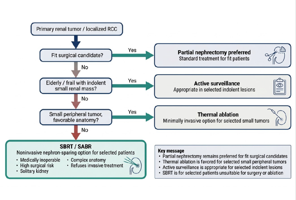
Figure 4. Proposed clinical positioning of SBRT among local treatment options for primary renal tumors. SBRT should not replace partial nephrectomy in suitable patients, but may serve as a noninvasive nephron-sparing option for selected patients unsuitable for surgery or thermal ablation.
In another meta-analysis of 13 prospective studies, overall local control for patients who underwent SBRT remained high at 98% at 1 year, 97% at 2 years, 95% at 3 years, with overall survival decreasing over time from 95% at 1 year, 86% at 2 years, and 78% at 3 years. Modest first-year eGFR reductions of -8.7 to -11 mL/min were also reported, as was low toxicity. The authors explicitly state that, despite these encouraging results, SBRT cannot yet be recommended routinely as an alternative to partial nephrectomy in candidates who would otherwise undergo surgery, due to the lack of randomized controlled trials (34).
However, in a recent systematic review by the International Society of Stereotactic Radiosurgery (ISRS), SBRT is recommended as an alternative for patients who are medically inoperable, at high surgical risk, or have declined surgery. The study showed a local control rate after BB of 94.1%, a five-year progression-free survival of 80.5%, and a five-year overall survival of 77.2%. The authors emphasize that the recommended doses are 25–26 Gy in 1 fraction for typical tumors and 42–48 Gy in 3 fractions for larger tumors. They also state that routine biopsy after SBRT is not recommended, as the above therapeutic approach is a safe and effective option for appropriate patients (6).
There is limited data comparing SBRT results with those observed with thermal ablation. One retrospective study examined OS after SBRT versus thermal ablation in patients with stage I RCC. In this analysis, SBRT was associated with worse OS than thermal ablation, with 3-year OS of 84% for cryoablation, 87% for RF/MW, and 76% for SBRT, and 5-year OS of 77% for cryoablation, 76% for RF/MW, and 58% for SBRT. It is also noted that patients with SBRT were generally older, more often female, with larger tumors, and more often in nonacademic centers. Overall, the authors conclude that SBRT for stage I RCC is associated with inferior OS compared with thermal ablation and suggest that it be used only in clinical trials or in exceptional cases until stronger comparative data are available (33).
A randomized trial was conducted in medically inoperable patients or those who refused surgery, comparing SBRT (25 Gy×1 fraction) with percutaneous RFA for renal masses ≤4 cm. In the first year of the study, renal functional preservation was similar across subgroups, with a mean change in eGFR of -3 mL/min with RFA versus -5.3 mL/min with SBRT (p=0.7). Safety was excellent, with no late toxicities reported in either arm. However, RFA demonstrated a higher rate of arterial enhancement loss than SBRT (83.3% versus 23%, p=0.041) and a higher pathologic response rate per protocol to the mandatory per-protocol biopsy. However, the authors report several technical weaknesses that need to be addressed, as well as the need for more long-term follow-up studies to provide a clearer picture of SBRT (35).
Thus, current evidence suggests that SBRT is not primarily a replacement for surgery in fit patients, but rather an important nephron-sparing noninvasive option for patients who are not ideal candidates for surgery or thermal ablation.
8. NCCN Guidance and Proposed Practical Recommendations
The National Comprehensive Cancer Network (NCCN) Kidney Cancer Guidelines recognizes radiation as a treatment option for selected patients with localized kidney cancers who are not candidates for surgery or other definitive local therapies (36). SBRT is the preferred radiation modality when radiation is utilized. For stage I disease (T1a and selected T1b tumors), SBRT carries a category 2A recommendation, indicating uniform NCCN consensus that the intervention is appropriate despite lower-level supporting evidence. For selected patients with stage II disease, SBRT is assigned a category 2B recommendation, reflecting NCCN consensus but less uniform agreement among panel members. This lack of consensus is evident later in the guideline document, where it notes that evidence supporting SBRT for tumors >7 cm (all stage II) remains limited. For stage III disease, SBRT carries a category 3 recommendation, indicating substantial disagreement among panel members regarding its appropriateness in this setting. Patient selection should incorporate tumor characteristics, patient fitness, and competing treatment options. Our approach to patient selection is depicted in Figure 4.
Partial or radical nephrectomy remains the preferred treatment for surgically eligible patients, while active surveillance may be appropriate for individuals with limited life expectancy or substantial frailty. SBRT is most appropriate for medically inoperable patients, those who decline surgery, or patients whose tumors are not suitable for thermal ablation due to size, location, or technical constraints. Additional considerations favoring SBRT include the desire to preserve renal function, avoid anesthesia-related risks, and pursue a noninvasive treatment approach.
The NCCN offers a reasonable follow-up strategy for patients treated with SBRT, which includes contrast-enhanced CT or MRI of the abdomen every 3 months for year 1, every 6 months for year 2, every 9 months for years 3-4, and annually in year 5. Because radiographic response following SBRT may be delayed, stable lesion size or persistent enhancement on early imaging should not be interpreted as treatment failure in the absence of progressive growth or other concerning findings. Renal function should be monitored longitudinally, particularly in patients with baseline chronic kidney disease or a solitary kidney.
9. Conclusion
The role of radiotherapy in the management of localized renal cell carcinoma has evolved substantially with the introduction of stereotactic body radiotherapy (SBRT). Advances in imaging, motion management, treatment planning, and image guidance have enabled the safe delivery of ablative radiation doses, overcoming many of the technical limitations that historically restricted the use of radiotherapy in RCC. Contemporary studies consistently demonstrate excellent local control, favorable toxicity profiles, and generally modest declines in renal function following treatment. However, the strongest evidence supporting SBRT exists for patients with smaller localized tumors who are medically inoperable, decline surgery, or are otherwise unsuitable candidates for thermal ablation. Data supporting its use in larger tumors and locally advanced disease remain more limited, and prospective studies are needed to better define outcomes in these populations.
Current evidence supports SBRT as an important noninvasive, nephron-sparing treatment option within the multidisciplinary management of localized RCC, but not as a replacement for surgery, in appropriately selected surgical candidates. Important questions remain regarding optimal patient selection, dose and fractionation strategies, long-term renal functional outcomes, and post-treatment response assessment. Further investigation is also needed to define the role of SBRT as both a definitive monotherapy and as a component of multimodality treatment strategies, including combinations with immunotherapy and targeted therapies. Continued prospective evaluation through collaborative efforts such as IROCK and future clinical trials will be essential to refine the role of SBRT and determine how best to integrate radiotherapy into the evolving treatment paradigm for RCC.
References
1. Gray RE, Harris GT. Renal Cell Carcinoma: Diagnosis and Management. Am Fam Physician. 2019;99(3):179-84.
2. Ali M, Ruiz VA, Psutka SP, Liu D, Siva S. 2022 WUOF/SIU International Consultation on Urological Diseases: Ablative Therapies for Localized Primary Renal Cell Carcinoma. Société Internationale d’Urologie Journal. 2022;3(6):437-49.
3. Richard PO, Violette PD, Bhindi B, Breau RH, Kassouf W, Lavallée LT, et al. Canadian Urological Association guideline: Management of small renal masses – Full-text. Can Urol Assoc J. 2022;16(2):E61-e75.
4. Wośkowiak P, Lewicka K, Bureta A, Salagierski M. Active surveillance and focal ablation for small renal masses: a better solution for comorbid patients. Arch Med Sci. 2020;16(5):1111-8.
5. Blanco AI, Teh BS, Amato RJ. Role of radiation therapy in the management of renal cell cancer. Cancers (Basel). 2011;3(4):4010-23.
6. Siva S, Louie AV, Kotecha R, Barber MN, Ali M, Zhang Z, et al. Stereotactic body radiotherapy for primary renal cell carcinoma: a systematic review and practice guideline from the International Society of Stereotactic Radiosurgery (ISRS). Lancet Oncol. 2024;25(1):e18-e28.
7. De Meerleer G, Khoo V, Escudier B, Joniau S, Bossi A, Ost P, et al. Radiotherapy for renal-cell carcinoma. Lancet Oncol. 2014;15(4):e170-7.
8. Siva S, Bressel M, Sidhom M, Sridharan S, Vanneste BGL, Davey R, et al. Stereotactic ablative body radiotherapy for primary kidney cancer (TROG 15.03 FASTRACK II): a non-randomised phase 2 trial. The Lancet Oncology. 2024;25(3):308-16.
9. Barbour AB, Upadhyay R, Anderson AC, Kutuk T, Kumar R, Wang SJ, et al. Stereotactic Body Radiation Therapy for Primary Renal Cell Carcinoma: A Case-Based Radiosurgery Society Practice Guide. Pract Radiat Oncol. 2025;15(1):74-85.
10. Suppression of homologydependent DNA doublestrand break repair induces PARP inhibitor sensitivity in VHLdeficient human renal cell carcinoma. Oncotarget.9(4).
11. Li B, Gao YJ, Wu XY, Cui J, Long Y, Xu JL, et al. Tumor-initiating cells contribute to radiation resistance in primary human renal clear cell carcinomas by activating the DNA damage checkpoint response. Oncol Lett. 2017;14(3):3261-7.
12. Yun E-J, Lin C-J, Dang A, Hernandez E, Guo J, Chen W-M, et al. Downregulation of Human DAB2IP Gene Expression in Renal Cell Carcinoma Results in Resistance to Ionizing Radiation. Clinical Cancer Research. 2019;25(14):4542-51.
13. Tselis N, Chatzikonstantinou G. Treating the Chameleon: Radiotherapy in the management of Renal Cell Cancer. Clin Transl Radiat Oncol. 2019;16:7-14.
14. Garcia-Barros M, Paris F, Cordon-Cardo C, Lyden D, Rafii S, Haimovitz-Friedman A, et al. Tumor response to radiotherapy regulated by endothelial cell apoptosis. Science. 2003;300(5622):1155-9.
15. Fuks Z, Kolesnick R. Engaging the vascular component of the tumor response. Cancer Cell. 2005;8(2):89-91.
16. Sonier M, Chu W, Lalani N, Erler D, Cheung P, Korol R. Evaluation of kidney motion and target localization in abdominal SBRT patients. J Appl Clin Med Phys. 2016;17(6):429-33.
17. Lo CH, Huang WY, Chao HL, Lin KT, Jen YM. Novel application of stereotactic ablative radiotherapy using CyberKnife(®) for early-stage renal cell carcinoma in patients with pre-existing chronic kidney disease: Initial clinical experiences. Oncol Lett. 2014;8(1):355-60.
18. Keller B, Bruynzeel AME, Tang C, Swaminath A, Kerkmeijer L, Chu W. Adaptive Magnetic Resonance-Guided Stereotactic Body Radiotherapy: The Next Step in the Treatment of Renal Cell Carcinoma. Front Oncol. 2021;11:634830.
19. Tetar SU, Bohoudi O, Senan S, Palacios MA, Oei SS, Wel AMV, et al. The Role of Daily Adaptive Stereotactic MR-Guided Radiotherapy for Renal Cell Cancer. Cancers (Basel). 2020;12(10).
20. Henke L, Kashani R, Robinson C, Curcuru A, DeWees T, Bradley J, et al. Phase I trial of stereotactic MR-guided online adaptive radiation therapy (SMART) for the treatment of oligometastatic or unresectable primary malignancies of the abdomen. Radiotherapy and Oncology. 2018;126(3):519-26.
21. Siva S, Ellis RJ, Ponsky L, Teh BS, Mahadevan A, Muacevic A, et al. Consensus statement from the International Radiosurgery Oncology Consortium for Kidney for primary renal cell carcinoma. Future Oncol. 2016;12(5):637-45.
22. Christensen M, Hannan R. The Emerging Role of Radiation Therapy in Renal Cell Carcinoma. Cancers (Basel). 2022;14(19).
23. Louie AV, Correa RJM, Boldt G, Zaorsky NG, Siva S. The Emerging Role of Stereotactic Radiotherapy in Primary Renal Cell Cancer: A Systematic Review of the Literature. International Journal of Radiation Oncology, Biology, Physics. 2019;105(1):E255-E6.
24. Grubb WR, Ponsky L, Lo SS, Kharouta M, Traughber B, Sandstrom K, et al. Final results of a dose escalation protocol of stereotactic body radiotherapy for poor surgical candidates with localized renal cell carcinoma. Radiotherapy and Oncology. 2021;155:138-43.
25. Siva S, Louie AV, Warner A, Muacevic A, Gandhidasan S, Ponsky L, et al. Pooled analysis of stereotactic ablative radiotherapy for primary renal cell carcinoma: A report from the International Radiosurgery Oncology Consortium for Kidney (IROCK). Cancer. 2018;124(5):934-42.
26. Grelier L, Baboudjian M, Gondran-Tellier B, Couderc A-L, McManus R, Deville J-L, et al. Stereotactic Body Radiotherapy for Frail Patients with Primary Renal Cell Carcinoma: Preliminary Results after 4 Years of Experience. Cancers. 2021;13(13):3129.
27. Pham D, Thompson A, Kron T, Foroudi F, Kolsky MS, Devereux T, et al. Stereotactic Ablative Body Radiation Therapy for Primary Kidney Cancer: A 3-Dimensional Conformal Technique Associated With Low Rates of Early Toxicity. International Journal of Radiation Oncology, Biology, Physics. 2014;90(5):1061-8.
28. Siva S, Bressel M, Sidhom M, Sridharan S, Vanneste B, Davey R, et al. TROG 15.03/ANZUP International Multicenter Phase II Trial of Focal Ablative STereotactic RAdiotherapy for Cancers of the Kidney (FASTRACK II). International Journal of Radiation Oncology, Biology, Physics. 2023;117(2):S3.
29. Siva S, Jackson P, Kron T, Bressel M, Lau E, Hofman M, et al. Impact of stereotactic radiotherapy on kidney function in primary renal cell carcinoma: Establishing a dose–response relationship. Radiotherapy and Oncology. 2016;118(3):540-6.
30. Ponsky L, Lo SS, Zhang Y, Schluchter M, Liu Y, Patel R, et al. Phase I dose-escalation study of stereotactic body radiotherapy (SBRT) for poor surgical candidates with localized renal cell carcinoma. Radiotherapy and Oncology. 2015;117(1):183-7.
31. Chang JH, Cheung P, Erler D, Sonier M, Korol R, Chu W. Stereotactic Ablative Body Radiotherapy for Primary Renal Cell Carcinoma in Non-surgical Candidates: Initial Clinical Experience. Clinical Oncology. 2016;28(9):e109-e14.
32. Tan VS, Correa RJM, Warner A, Ali M, Muacevic A, Ponsky L, et al. Long-term Renal Function Outcomes After Stereotactic Ablative Body Radiotherapy for Primary Renal Cell Carcinoma Including Patients with a Solitary Kidney: A Report from the International Radiosurgery Oncology Consortium of the Kidney. Eur Urol Oncol. 2024;7(6):1527-34.
33. Uhlig A, Uhlig J, Trojan L, Kim HS. Stereotactic Body Radiotherapy for Stage I Renal Cell Carcinoma: National Treatment Trends and Outcomes Compared to Partial Nephrectomy and Thermal Ablation. Journal of Vascular and Interventional Radiology. 2020;31(4):564-71.
34. Suleja A, Bilski M, Laukhtina E, Fazekas T, Matsukawa A, Tsuboi I, et al. Stereotactic Body Radiotherapy (SBRT) for the Treatment of Primary Localized Renal Cell Carcinoma: A Systematic Review and Meta-Analysis. Cancers. 2024;16(19):3276.
35. Swaminath A, Cassim R, Millan B, Mironov O, Ahir P, Tajzler C, et al. Final Results from a Prospective Randomized Pilot Trial of Stereotactic Body Radiation Therapy vs. Radiofrequency Ablation for the Management of Small Renal Masses (RADSTER). International Journal of Radiation Oncology, Biology, Physics. 2023;117(2):S82.
36. Network® NCC. NCCN Clinical Practice Guidlines in Oncology (NCCN Guidelines®) – Kidney cancer 2026 [updated April 14, 2026. Version 2.2026:[Available from: https://www.nccn.org/professionals/physician_gls/pdf/kidney.pdf.