#Equal Contributions as Senior/Supervising Authors
ϮAddress Correspondence:
Abhishek A. Chakraborty, PhD
Department of Cancer Sciences, Cleveland Clinic, Cleveland OH
E-mail: chakraa@ccf.org
Conflict of Interest: The authors declare no conflict of interest with this study.
INTRODUCTION
Clinical outcomes for patients diagnosed with renal cell carcinoma (RCC) have dramatically improved over the last few decades and the identification of the best management strategies are now being actively debated [1]. In particular, the approval of immunotherapy (IO) for adjuvant RCC and IO-based combinations in patients with metastatic RCC (mRCC) have improved overall survival (OS) [2-5]. However, despite these improvements, biomarkers for localized versus mRCC as well as biomarkers of OS are severely lacking. Currently available algorithms estimating the risk of RCC recurrence after surgery for localized RCC, as well as prognostic scoring models for patients with mRCC, are primarily comprised of clinical features and do not account for the heterogeneity of underlying disease biology [6-9]. The need for novel biologically-driven biomarkers is thus critical.
Metastatic kidney cancer presents unique biological challenges in the development of circulating biomarkers. Although there has been some success in using cell-free diagnostic tools, kidney cancers do not shed biological material efficiently, which has limited the utility of these approaches in biomarker discovery [10]. The development of tumors in the kidney disrupts glomerular architecture and filtration and thereby offers an opportunity to leverage modern technologies to diagnose this filtration defect in mRCC patients. Plasma Kidney Injury Molecule 1 (KIM-1) represents one such recent example of a circulating biomarker of nephric damage that is being extensively exploited for annotation of disease stage and treatment outcomes [11 12].
The development of the multiplexed Olink® proteomics platform approach has offered new technology for biomarker discovery in many human disease contexts [13]. The discovery of KIM-1 as a biomarker of therapy response in RCC was made using such multiplexed proteomics tools [12]. Olink® uses a ‘proximity extension assay’ where the same target protein is recognized using two independent antibodies, each with a conjugated complementary DNA tail [14]. The proximal association of the two antibodies can be quantified using sequencing-based strategies. The technology has now matured, and commercial Olink® panels now cater to specific functionally annotated protein target panels (e.g., metabolic panels, immunological panels, etc.). These focused approaches have reduced biological noise and improved biomarker discovery.
Gut microbial dysbiosis and the use of microbial agents as adjuvants are both relevant determinants of immunotherapy response in kidney cancer [15-17]. The nephric damage induced by RCC lesions, especially in the proximal tubules, compromises estimated Glomerular Filtration Rates (eGFR). One unexpected consequence of this disruption is the re-entry of gut microbiome derived metabolites into circulation. Improved understanding of gut microbial composition and their metabolic products, in concert with the development of highly sensitive, targeted mass spectrometry approaches has led to exponential leaps in our ability to utilize microbial metabolites as biomarkers in human disease. However, these technologies have thus far not been leveraged for biomarker discovery in mRCC.
In this study we objectively evaluate our ability to use these recent technological advances for biomarker discovery in mRCC. Remarkably, our studies have revalidated soluble PD-L1 (sPD-L1) as a biomarker of disease progression and led to the identification of several novel protein and microbial metabolite biomarkers that are elevated in mRCC patients. Finally, we have also identified prognostic biomarkers that are associated with better overall survival in these patients.
RESULTS
Cohort Characteristics
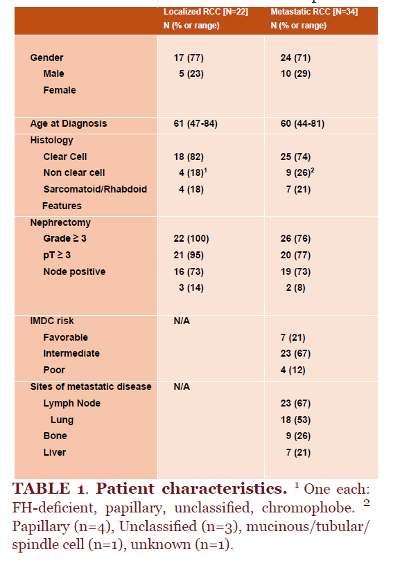
A total of 56 patients with RCC who had at least one blood sample available for analysis were included (Table 1). Of these patients, 22 had localized RCC and 34 had mRCC. Of the patients with localized disease, most were male (77%) with a median age of 61 (range, 47-84). All patients with localized RCC underwent a nephrectomy as primary treatment for their disease with clear cell RCC (82%) as the most common histology. Patients with metastatic disease were mostly male (71%), median age of 60 (range, 44-81), with the majority having undergone a prior nephrectomy (76%). Other clinical features including histology and metastatic sites of disease were reflective of a typical mRCC cohort (Table 1). The median OS for patients with localized disease was not reached (NA-NA) and for patients with mRCC was 44.32 months (35.84-NA; p=0.005).
Circulating protein biomarkers in mRCC
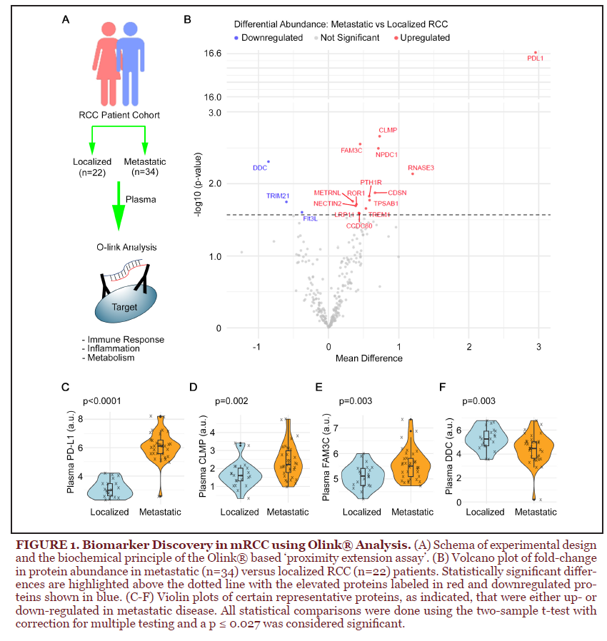
We analyzed banked plasma samples from the RCC cohort using three Olink® T96 panels: immune response, inflammation, and metabolism (Figure 1A and Supplementary Table 1), altogether representing ~276 proteins (92 unique protein probes in each panel). The circulating levels of these proteins were measured using the standard manufacturer recommended protocol; however, to minimize bias, localized and mRCC samples were randomized and then added to the same plate. Proteins were quantified and the difference in circulating levels was compared using a two-sample t-test (adjusted p ≤ 0.027). The differences in circulating levels of the proteins in the two cohorts were visualized using a volcano plot and showed 13 upregulated and 3 downregulated proteins in the mRCC cohort (Figure 1B).
We noted significant biological value in these results. Most notably, we observed that circulating levels of sPD-L1 – a regulator of immune checkpoint response – were elevated in mRCC patients (Figure 1C). This is consistent with a previous report showing sPD-L1 to be a biomarker of progressive disease in RCC [18]. Other notable proteins that were elevated in mRCC included the cell state regulatory proteins [e.g., Meteorin like glial cell differentiation regulator (METRNL), Nectin Cell Adhesion Molecule 2 (NECTIN2), and Neuronal Proliferation and Differentiation Control 1 (NPDC1)] and signaling receptors [e.g., LDL Receptor related Protein 11 (LRP11), Receptor tyrosine kinase like orphan receptor 1 (ROR1), and FAM3 Metabolism Regulating Signaling Molecule C (FAM3C)] (Figures 1B to 1F and Supplementary Table 2). The elevated expression of these proteins is consistent with the epithelial-mesenchymal transition in mRCC and the potential immune checkpoint activation in these cancers. In contrast, we found that the circulating levels of Dopamine Decarboxylase (DDC) and Tripartite Motif-Containing Protein 21 (TRIM21) were reduced in mRCC patients. Altogether, this analysis offered many candidate biomarkers of metastatic disease.
Prognostic markers in mRCC
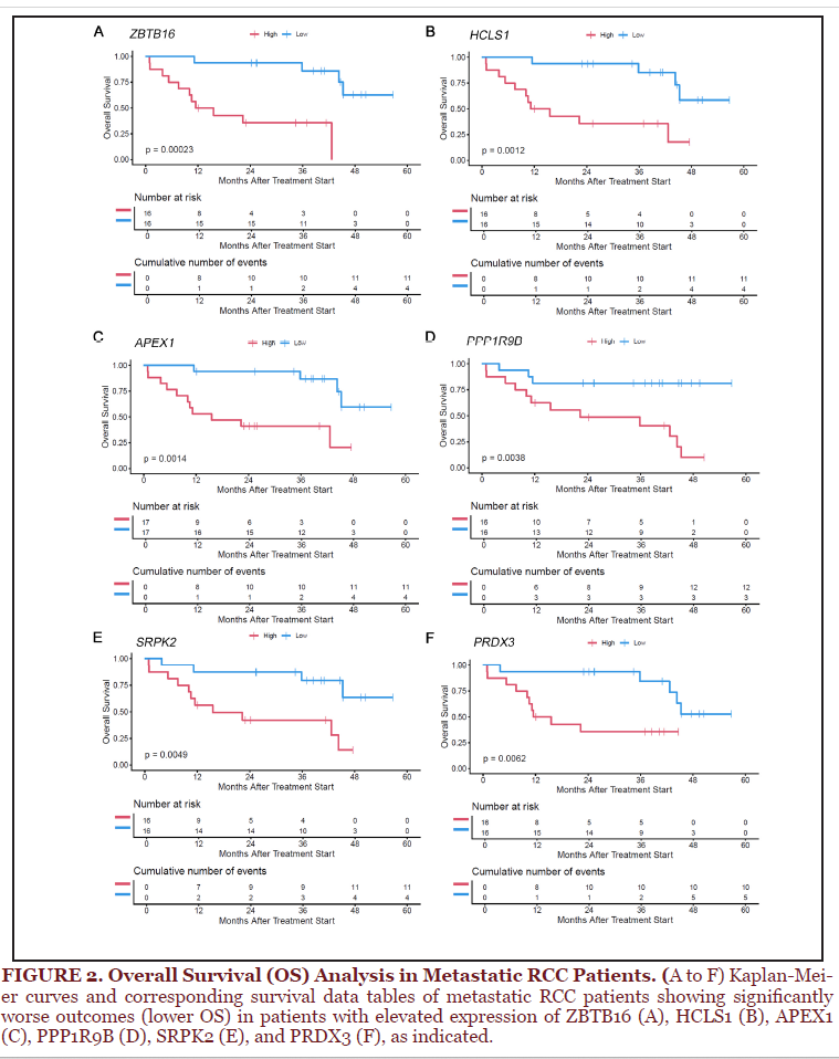
As expected, we noted that patients with mRCC had worse overall survival (OS) than patients with localized disease (Supplementary Figure 1). We then re-analyzed the metastatic cohort and probed for prognostic markers in the Olink® panel. We stratified the mRCC patients into the top (high) or bottom (low) median of protein expression and compared the association of protein expression with OS. The results of this analysis were surprising. For example, none of the proteins with elevated circulating levels in the mRCC patients (relative to localized RCC) showed any statistically significant associations with OS within the mRCC patient cohort. Instead, we found ~12 proteins whose higher circulating levels correlated with worse OS in mRCC (Supplementary Table 3). Besides the well-known oncogenic transcription factor, JUN, we identified transcriptional regulatory proteins [e.g., HEXIM P-TEFb complex subunit 1 (HEXIM1) and Zinc Finger and BTB domain containing 16 (ZBTB16)] and signaling proteins [e.g., Hematopoietic cell-specific Lyn substrate 1 (HCLS1) and SRSF protein kinase 2 (SPRPK2)] among the proteins associated with worse outcomes in mRCC (Figure 2 and Supplementary Table 3). Note that circulating levels of these proteins were not increased in mRCC relative to localized disease, thus highlighting the need to distinguish expression levels from potential functional consequences of these proteins. Altogether, our Olink® analysis was able to identify new biomarkers associated with mRCC disease.
Discovery of differentially abundant microbial metabolites
One concern with the Olink® biomarkers was that, except for sPD-L1, the other markers showed relatively small fold-change differences in metastatic versus localized disease. We therefore sought to leverage our expertise to interrogate other novel biological processes, such as the gut microbial metabolites, for biomarker discovery.
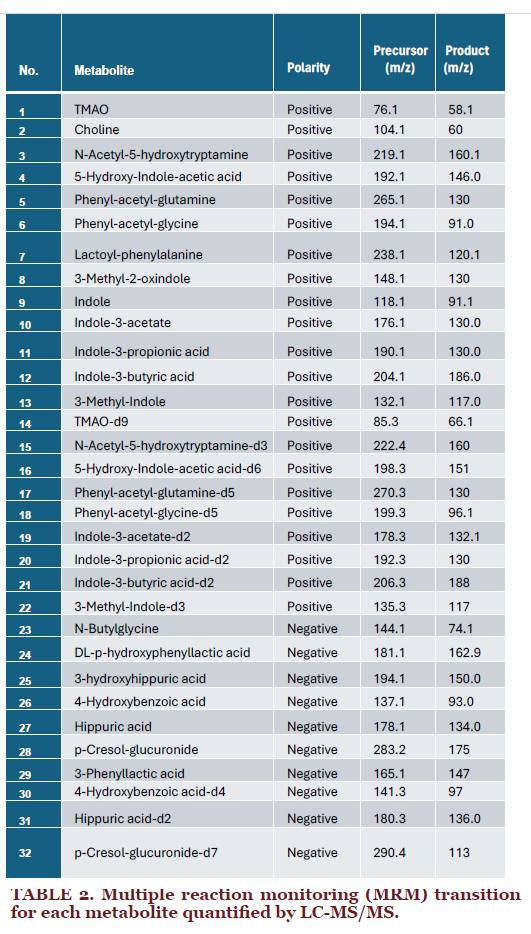
The gut microbial community synthesizes and catabolizes many key nutrients and in fact can be considered an endocrine organ producing unique metabolites that systemically enter circulation [19 20]. Moreover, gut dysbiosis is a clinically relevant feature of kidney cancer [15-17]. We hypothesized that the disruption of the kidney architecture in RCC patients impacts the circulating levels of many of these metabolites and interrogated the relevance of these metabolites as mRCC biomarkers. To this end, we deployed a targeted stable isotope dilution liquid chromatography tandem mass spectrometry (LC-MS/MS) method to measure the abundance of a panel of circulating metabolites that are known to originate from gut bacteria (Figure 3A and Table 2). We then compared the circulating levels of metabolites derived from indole and choline in the plasma of mRCC (versus localized RCC) patients, using the same statistical parameters described for the proteomics analysis (two-sample t-test, adjusted p-value ≤ 0.027). These comparisons identified three metabolites whose circulating levels were elevated in mRCC patients, including bacterially-derived metabolites of tryptophan, such as indole (fold-change: 4.1; p-value: 1.51E-06) and 5-hydroxy-indole acetic acid (5-HIAA, fold-change: 1.7; p-value: 2.42E-03) (Figure 3B and 3C, and Supplementary Table 2). We also found that levels of the bacterial phenylalanine metabolite phenylacetylglutamine (PAGln) were elevated in metastatic patients (fold-change: 2.3; p-value: 4.90E-03) (Figure 3D and Supplementary Table 2).
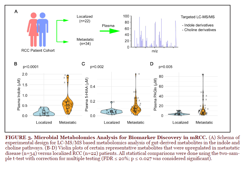
The discovery of indole derivatives and PAGln as biomarkers of mRCC deserves further investigation, given the emerging literature supporting a role for the gut microbiome in ccRCC [21-23]. The indole metabolites are derived from tryptophan; however, their effects on tumor growth have been controversial [24 25]. Plant-derived indole metabolites (e.g., the indole alkaloids Vinblastine and Vinchristine and indole-3-carbinol), are known for their anti-tumor properties [26]. Interestingly, indole is metabolized by gut microbes into indoxyl sulfate, a uremic toxin associated with chronic renal damage [27]. Moreover, studies in mRCC patients indicate that indole (and its derivatives) are associated with immunosuppression and driving metastatic disease [28]. These findings are consistent with the idea that elevated circulating levels of indole in patients with mRCC might be associated with dysfunctional tumor-immune interactions and ultimately promote tumor growth in kidney cancer.
In contrast to the indole derivatives, the relevance of PAGln is relatively understudied in cancer biology; however, this molecule is associated with worse cardiovascular risk and with renal dysfunction, which has led to its previous characterization as a uremic toxin [29-33]. Therefore, elevated PAGln (like indoxyl sulfate) could potentially be a marker of renal damage in mRCC patients.
Correlation analysis of the Olink® and microbial metabolite panels
Our initial studies analyzed the Olink® and microbial metabolite panels independently to identify novel biomarkers in mRCC patients. Next, we performed correlation analysis between these two datasets to identify biological features that co-occur with high propensity. To perform this analysis, we did pairwise comparisons between the microbial metabolites with each of the proteins in the Olink® panel. We calculated statistical significance using the Spearman’s correlation test (FDR: 10%, adjusted p-value ≤ 0.0482). Our analysis identified ~735 protein-metabolite pairs that showed statistically significant correlations (Supplementary Figure 2 and Supplementary Table 4).
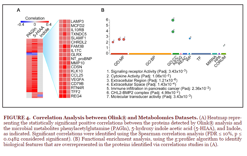
We then focused on the three microbial metabolites (Indole, 5-HIAA, and PAGln) that showed significant elevation in mRCC patient samples and shortlisted the statistically significant correlations for each of these metabolites (Figure 4A and Supplementary Table 4). All three metabolites correlated with a panel of ~20 proteins (Figure 4A). To infer the biological implications of these associations, we performed enrichment analysis on the proteins that showed statistically significant correlations with these metabolites, using the g:profiler algorithm [34]. These studies demonstrated that the three metabolites correlated with elevated circulating levels of proteins involved in chemokine signaling, receptor-ligand signaling modules, and chemotaxis/migration (Figure 4B), consistent with the immunological dysregulation and inflammatory state that is typical of metastatic tumors.
DISCUSSION
Renal cell carcinoma (RCC) is one of the most common cancers in the United States with approximately 80,000 new diagnoses per year and 14,000 deaths from RCC each year [35]. Despite the high prevalence and lethality of RCC, there are no established blood-based biomarkers for early detection or treatment selection. The International Metastatic Renal Cell Carcinoma Database Consortium (IMDC) is the most commonly used prognostic criteria currently used in clinical practice [7 36], but it relies solely on clinical characteristics. It does not account for biological heterogeneity, such as underlying genomic, metabolomic, or microbial differences between RCC patients. While it functions well as a prognostic marker for mRCC patients undergoing systemic therapy, it does not guide treatment selection or serve as a predictor of response or resistance to therapy. Other algorithms in RCC are also predominantly limited to clinical features.
Recently, investigations into the genomic heterogeneity of RCC and an attempt to incorporate their use in prognostication and treatment selection has gained momentum. Efforts to incorporate genomic components (e.g., PBRM1, BAP1, etc.) into the IMDC prognostic criteria are ongoing. Studies such as OPTIC (NCT05361720), BIONIKK (NCT02960906), and CARE1 (NCT06364631) are using genomic biomarkers to guide treatment selection. Moreover, studies investigating the role of blood-based biomarkers such as KIM-1 to detect RCC before clinical diagnosis or as a means to monitor effective therapeutic response are also underway [11 12]. Despite this progress, no validated biomarkers are currently available, highlighting the need for novel approaches in biomarker development for RCC. Our study provides a template to complement these ongoing trials with biomarker discovery by leveraging some recent technological developments.
Although largescale biomarker discovery was initially limited to nucleic acids, recent technological developments have allowed us to directly measure small molecules and proteins. The development of Olink® has been one such example. This multiplexed assay relies on proximity ligation. Two monoclonal antibodies targeting different epitopes on the same protein, each with a complementary DNA tail, are bound to biological samples. The juxtaposition of the antibodies results in DNA hybridization, which can be efficiently quantified using realtime-qPCR analysis. The abundance of the hybridized product thus offers a quantitative proxy for binding affinity. This technological approach has now matured and begun offering the opportunity for largescale biomarker discovery. Our work has leveraged this relatively novel tool to address the clinically urgent need for prognostic and therapeutic biomarker discovery in mRCC.
Within the last decade, there have been numerous reports linking the gut microbiome and metaorganismal metabolic pathways to diverse cancers [37-39]. However, the vast majority of cancer-related microbiome studies are correlative, simply cataloguing stool microbiome or tumor-associated microbiome in cancer patients using metagenomic sequencing approaches. Undoubtedly, high throughput sequencing approaches have uncovered a number of compelling microbiome-cancer links. However, just because a bacterial sequence is detectable and is associated with disease does not mean that there is a functional alteration in the gut microbiome. In parallel to the rapid growth in sequencing-based approaches, advances in the fields of mass spectrometry and computational biology have allowed for the identification of natural products and metabolites that originate solely from the gut microbial endocrine organ [19 20]. Here, we have leveraged access to LC-MS/MS methods to quantify several metabolites that are known to be exclusively derived from bacterial sources. Although, here, we have only established associations between the metabolites and mRCC, these findings could be mechanistically developed to address the importance of microbe-host crosstalk.
There is now a wealth of evidence supporting a role for metaorganismal tryptophan metabolism in diverse cancers. In general, the host derived tryptophan metabolite kynurenine has been linked to cancer promotion [40-42], whereas gut microbe-derived indole metabolites of tryptophan such as indole-3-propionic acid and indole-3-acetic acid have been shown to enhance anti-tumor immunity [40-42]. The current thought is that bacterially-derived indole metabolites are sensed by the host nuclear hormone receptor aryl hydrocarbon receptor (AhR) to elicit some of their biological activity in the host [43 44]. However, we still incompletely understand potential mechanisms by which microbial and host-derived tryptophan metabolites impact ccRCC metastasis. Interestingly, both AhR and the Hypoxia Inducible Factor (HIF) transcription factor compete for binding with the Aryl Hydrocarbon receptor Nuclear Translocator (ARNT, also called HIF1β). HIF is a well-known oncogenic driver in ccRCC and we speculate that indole-derived metabolites could thus functionally impact HIF-dependent oncogenic programs by regulating the available intracellular pools of ARNT. Altogether, additional studies are warranted to explore the potential role for bacterially-derived indole metabolites in ccRCC progression.
Our data also indicates elevated levels of the gut microbe-derived phenylalanine metabolite PAGln in mRCC patients. PAGln has recently gained attention as a cardiovascular disease-associated metabolite, where it is known to signal through host adrenergic receptors to promote pro-thrombotic effects [29-33]. We thus speculate that gut microbe-derived PAGln may similarly activate adrenergic pathways in the kidney or metastatic niches to promote ccRCC. Collectively, the current study identifies several potential metaorganismal nutrient metabolism pathways (tryptophan > indole; phenylalanine > PAGln) that deserve further mechanistic investigation in the setting of ccRCC metastasis. Although both these metabolites have associations with nephric integrity, we did not have the clinical measures of renal function from our patients, and thus could not statistically correlate circulating levels of these metabolites with renal damage.
There are several inherent limitations in this study. Given the relative expense associated with these analyses, we have presently limited our studies to a pilot ‘risk-assessment’ scale with 56 patient samples, which has unfortunately diminished our ability to make broader associations with disease outcomes. Moreover, clinically, the study contains a relatively small and heterogeneous population of patients with mRCC and does not define for which specific histology, stage, or systemic treatment these findings are likely to be most relevant. Another important limitation is that there were no standardized collection time points for these samples. As such, timing of plasma collection relative to nephrectomy, metastatic diagnosis, etc. varied between patients and may have impacted results. Finally, the study is also conducted at a single institution and thus requires external validation in larger cohorts. Importantly, sPD-L1 was the most notable biomarker identified in our Olink® analysis and circulating levels of this protein (previously called soluble PD-L1) have been reported as a biomarker of RCC disease progression [18]. Thus, while additional studies are indeed necessary, our preliminary findings do have some external validation and thus bear potential for future clinical translation.
Finally, it is critical to highlight that the metastatic biomarkers (i.e., those elevated in mRCC vs localized RCC) did not translate into an association with OS within mRCC patients. In other words, biomarkers of a disease state are not necessarily prognostic biomarkers within a specific disease setting. We acknowledge the caveat that the abundance of a given protein and its prognostic association with OS neither establishes causation nor offers any mechanistic links between the protein (and/or metabolite) and mRCC development. Expression levels and prognostic associations are routinely – and erroneously – conflated into evidence of causality and therapeutic feasibility [45]. Instead, we cautiously offer these differentially regulated proteins and metabolites purely as biomarkers of metastatic disease. Future studies will address the functional relevance of these proteins/metabolites as (potential) drivers of metastatic disease by leveraging cell-based assays, animal models of kidney cancer, and human patient-derived organoids/xenografts. Altogether, the biomarkers identified in this study have provided us with important results that serve as a foundation for future clinical and laboratory-based validation studies.
ACKNOWLEDGMENTS
The authors declare no conflicts of interest with the findings presented in this work. All data is provided in the manuscript or as supplementary data. The following author contributions are credited. Conceptualization: MCO, JMB, AAC; Performed Experiments: XY, SD, VV, MP; Experimental Data Analysis: XY, SD, VV, ML, MP, CMDM; Statistical Analysis: WW; Wrote Manuscript: MCO, JMB, AAC.
FUNDING
This work was funded by a DOD-KCRP-TRPA grant (W81XWH2210430; PI: MCO and JMB; Co-I: AAC).
DATA AVAILABILITY STATEMENT
All the data necessary to evaluate the conclusions of the manuscript are provided in the paper and/or the Supplementary Materials.
METHODS
IRBs and Patient consent details
All plasma samples were collected upon consent from kidney cancer patients at the Cleveland Clinic under the institutionally approved protocols (IRB4639, CASE5816, and CASE12815), which were combined into a single standalone IRB 22-676 to support this DOD funded study.
STATISTICAL METHODS
Baseline biomarker expressions were summarized using mean and SD by disease group. Two-sample t-test was used to compare each biomarker’s expression between disease groups. False discovery rate (FDR) control was performed using beta-uniform method with 20% FDR rate. Spearman’s correlation test was used to correlate metabolite biomarkers with Olink® biomarkers. Due to a large number of significant correlation p-values, FDR was set more stringently at 10% to identify significantly correlated biomarker pairs. Overall survival (OS) was estimated by the Kaplan-Meier method. Log rank test was used to compare OS between disease groups and biomarkers, where biomarkers were dichotomized at median and the FDR of the log rank test p-values was set at 20%. All tests were two-sided and p-values passing FDR thresholds were considered statistically significant. Statistical analysis was carried out using SAS Studio 3.7 (SAS Institute, Cary, NC) and R version 4.4 (R Foundation, Vienna, Austria).
Olink® Analysis
The Olink® analysis was performed on three panels (described in Supplementary Table 1). The patient plasma samples were first randomized and the users were blinded to the sample identity. The localized and mRCC samples were spread uniformly (and randomly) on the analysis plates. All assays were done using 1 μl of the plasma, which was aliquoted by thawing frozen plasma samples. All prepared sample plates were held overnight at -80°C until the day of experiment. The analysis was done by incubating samples with paired antibodies linked to DNA oligonucleotides for 16-22 hours. Enzymatic extension was performed and the new (hybridized) DNA sequences were amplified and quantified using the Q100 instrument. Each plate underwent quality control analysis by the immune monitoring laboratory’s technical staff. Only plates passing quality control were analyzed and compiled into final assay results.
LC-MS/MS based targeted metabolomics
Stable isotope dilution liquid chromatography with quadrupole mass spectrometer (LC-
MS/MS) was used for quantification of gut microbiome-associated metabolites in plasma. 20 µl of plasma was used for extraction. In brief, 80 µl of isotope labeled internal standard mix was added to 20 µl of plasma, vortexed for 1 min, followed by centrifuging at 20000 g at 4 ºC for 15 min. 30 µl of supernatant was transferred to a MS vial with insert, then 30 ul of LCMS water was added to the MS vial for the final analysis. Plasma samples were analyzed on a Thermo Vanquish liquid chromatograph coupled with a Thermo TSQ Quantiva. 0.2% of formic acid in LC-MS water was used as mobile phase A and 0.2% formic acid in acetonitrile was used as mobile phase B. The separation was conducted using the following gradient: 0 min, 5% B; 0-2 min, 5% B; 2-8 min, 5%-100%B; 8-16 mins,100%B; 16-16.5 mins, 5%B; 16.5-25 mins, 5%B. The flow rate was set at 0.2 ml/min. Samples were injected at 2 μl onto a Phenomenex Gemini C18 analytical column (2.0×150 mm, 3 μm). Column temperature was set at 25 °C. Multiple reaction monitoring (MRM) ion transitions are shown in Table 2. The acquired data was processed by Thermo Xcalibur 4.3 software to calculate the concentrations.
REFERENCES
1. McKay RR, Pal S, Xie W, et al. Advanced Urologic Cancer Consensus Conference (AUC3) 2025: Expert consensus on the management of renal cell and urinary tract cancers. CA Cancer J Clin 2026;76(1):e70052 doi: 10.3322/caac.70052.
2. Barragan-Carrillo R, Saad E, Saliby RM, et al. First and Second-line Treatments in Metastatic Renal Cell Carcinoma. Eur Urol 2025;87(2):143-54 doi: 10.1016/j.eururo.2024.10.019 [published Online First: 20241106].
3. Choueiri TK, Tomczak P, Park SH, et al. Overall Survival with Adjuvant Pembrolizumab in Renal-Cell Carcinoma. N Engl J Med 2024;390(15):1359-71 doi: 10.1056/NEJMoa2312695.
4. Motzer R, Alekseev B, Rha SY, et al. Lenvatinib plus Pembrolizumab or Everolimus for Advanced Renal Cell Carcinoma. N Engl J Med 2021;384(14):1289-300 doi: 10.1056/NEJMoa2035716 [published Online First: 20210213].
5. Motzer RJ, Tannir NM, McDermott DF, et al. Nivolumab plus Ipilimumab versus Sunitinib in Advanced Renal-Cell Carcinoma. N Engl J Med 2018;378(14):1277-90 doi: 10.1056/NEJMoa1712126 [published Online First: 20180321].
6. Correa AF, Jegede OA, Haas NB, et al. Predicting Disease Recurrence, Early Progression, and Overall Survival Following Surgical Resection for High-risk Localized and Locally Advanced Renal Cell Carcinoma. Eur Urol 2021;80(1):20-31 doi: 10.1016/j.eururo.2021.02.025 [published Online First: 20210309].
7. Heng DY, Xie W, Regan MM, et al. Prognostic factors for overall survival in patients with metastatic renal cell carcinoma treated with vascular endothelial growth factor-targeted agents: results from a large, multicenter study. J Clin Oncol 2009;27(34):5794-9 doi: 10.1200/JCO.2008.21.4809 [published Online First: 20091013].
8. Leibovich BC, Blute ML, Cheville JC, et al. Prediction of progression after radical nephrectomy for patients with clear cell renal cell carcinoma: a stratification tool for prospective clinical trials. Cancer 2003;97(7):1663-71 doi: 10.1002/cncr.11234.
9. Motzer RJ, Mazumdar M, Bacik J, Berg W, Amsterdam A, Ferrara J. Survival and prognostic stratification of 670 patients with advanced renal cell carcinoma. J Clin Oncol 1999;17(8):2530-40 doi: 10.1200/JCO.1999.17.8.2530.
10. Zengin ZB, Weipert C, Salgia NJ, et al. Complementary Role of Circulating Tumor DNA Assessment and Tissue Genomic Profiling in Metastatic Renal Cell Carcinoma. Clinical cancer research : an official journal of the American Association for Cancer Research 2021;27(17):4807-13 doi: 10.1158/1078-0432.CCR-21-0572 [published Online First: 20210615].
11. Xu W, Gaborieau V, Niman SM, et al. Plasma Kidney Injury Molecule-1 for Preoperative Prediction of Renal Cell Carcinoma Versus Benign Renal Masses, and Association With Clinical Outcomes. J Clin Oncol 2024;42(22):2691-701 doi: 10.1200/JCO.23.00699 [published Online First: 20240503].
12. Rini BI, Albiges L, Tang X, et al. Circulating kidney injury molecule-1 (KIM-1) and association with outcome to adjuvant immunotherapy in renal cell carcinoma. Ann Oncol 2025;36(12):1525-34 doi: 10.1016/j.annonc.2025.08.007 [published Online First: 20250828].
13. Alvez MB, Bergstrom S, Kenrick J, et al. A human pan-disease blood atlas of the circulating proteome. Science 2025;390(6779):eadx2678 doi: 10.1126/science.adx2678 [published Online First: 20251218].
14. Assarsson E, Lundberg M, Holmquist G, et al. Homogenous 96-plex PEA immunoassay exhibiting high sensitivity, specificity, and excellent scalability. PloS one 2014;9(4):e95192 doi: 10.1371/journal.pone.0095192 [published Online First: 20140422].
15. Dizman N, Meza L, Bergerot P, et al. Nivolumab plus ipilimumab with or without live bacterial supplementation in metastatic renal cell carcinoma: a randomized phase 1 trial. Nature medicine 2022;28(4):704-12 doi: 10.1038/s41591-022-01694-6 [published Online First: 20220228].
16. Dizman N, Hsu J, Bergerot PG, et al. Randomized trial assessing impact of probiotic supplementation on gut microbiome and clinical outcome from targeted therapy in metastatic renal cell carcinoma. Cancer Med 2021;10(1):79-86 doi: 10.1002/cam4.3569 [published Online First: 20201101].
17. Routy B, Le Chatelier E, Derosa L, et al. Gut microbiome influences efficacy of PD-1-based immunotherapy against epithelial tumors. Science 2018;359(6371):91-97 doi: 10.1126/science.aan3706 [published Online First: 20171102].
18. Mahoney KM, Ross-Macdonald P, Yuan L, et al. Soluble PD-L1 as an early marker of progressive disease on nivolumab. J Immunother Cancer 2022;10(2) doi: 10.1136/jitc-2021-003527.
19. Brown JM, Hazen SL. The gut microbial endocrine organ: bacterially derived signals driving cardiometabolic diseases. Annu Rev Med 2015;66:343-59 doi: 10.1146/annurev-med-060513-093205.
20. Massey W, Brown JM. The Gut Microbial Endocrine Organ in Type 2 Diabetes. Endocrinology 2021;162(2) doi: 10.1210/endocr/bqaa235.
21. Yang BY, Zhao FZ, Li XH, et al. Alteration of pro-carcinogenic gut microbiota is associated with clear cell renal cell carcinoma tumorigenesis. Front Microbiol 2023;14:1133782 doi: 10.3389/fmicb.2023.1133782 [published Online First: 20230405].
22. Chen Y, Ma J, Dong Y, et al. Characteristics of Gut Microbiota in Patients With Clear Cell Renal Cell Carcinoma. Front Microbiol 2022;13:913718 doi: 10.3389/fmicb.2022.913718 [published Online First: 20220704].
23. Liss MA, Chen Y, Rodriguez R, et al. Microbiome within Primary Tumor Tissue from Renal Cell Carcinoma May Be Associated with PD-L1 Expression of the Venous Tumor Thrombus. Adv Urol 2020;2020:9068068 doi: 10.1155/2020/9068068 [published Online First: 20200218].
24. Yan J, Chen D, Ye Z, et al. Molecular mechanisms and therapeutic significance of Tryptophan Metabolism and signaling in cancer. Mol Cancer 2024;23(1):241 doi: 10.1186/s12943-024-02164-y [published Online First: 20241030].
25. Hou Y, Li J, Ying S. Tryptophan Metabolism and Gut Microbiota: A Novel Regulatory Axis Integrating the Microbiome, Immunity, and Cancer. Metabolites 2023;13(11) doi: 10.3390/metabo13111166 [published Online First: 20231120].
26. Hassan SM, Farid A, Panda SS, et al. Indole Compounds in Oncology: Therapeutic Potential and Mechanistic Insights. Pharmaceuticals (Basel) 2024;17(7) doi: 10.3390/ph17070922 [published Online First: 20240710].
27. Rattanasompattikul M, Srithongkul T, Tantisattamo E, Kalantar-Zadeh K, Noppakun K. Indoxyl sulfate: clinical implications for anemia management in chronic kidney disease. Curr Opin Nephrol Hypertens 2025 doi: 10.1097/MNH.0000000000001145 [published Online First: 20251208].
28. Bari S, Manley BJ, Muzaffar J, et al. Association of microbial metabolism of tryptophan with resistance to immune checkpoint (ICB) therapy in renal cell cancer (RCC). Journal of Clinical Oncology 2024;42(4_suppl):452-52 doi: 10.1200/JCO.2024.42.4_suppl.452.
29. Nemet I, Saha PP, Gupta N, et al. A Cardiovascular Disease-Linked Gut Microbial Metabolite Acts via Adrenergic Receptors. Cell 2020;180(5):862-77 e22 doi: 10.1016/j.cell.2020.02.016.
30. Hobson S, Qureshi AR, Ripswedan J, et al. Phenylacetylglutamine and trimethylamine N-oxide: Two uremic players, different actions. Eur J Clin Invest 2023;53(12):e14074 doi: 10.1111/eci.14074 [published Online First: 20230807].
31. Nemet I, Li XS, Haghikia A, et al. Atlas of gut microbe-derived products from aromatic amino acids and risk of cardiovascular morbidity and mortality. Eur Heart J 2023;44(32):3085-96 doi: 10.1093/eurheartj/ehad333.
32. Romano KA, Nemet I, Prasad Saha P, et al. Gut Microbiota-Generated Phenylacetylglutamine and Heart Failure. Circ Heart Fail 2023;16(1):e009972 doi: 10.1161/CIRCHEARTFAILURE.122.009972 [published Online First: 20221216].
33. Zhu Y, Dwidar M, Nemet I, et al. Two distinct gut microbial pathways contribute to meta-organismal production of phenylacetylglutamine with links to cardiovascular disease. Cell Host Microbe 2023;31(1):18-32 e9 doi: 10.1016/j.chom.2022.11.015 [published Online First: 20221221].
34. Kolberg L, Raudvere U, Kuzmin I, Adler P, Vilo J, Peterson H. g:Profiler-interoperable web service for functional enrichment analysis and gene identifier mapping (2023 update). Nucleic acids research 2023;51(W1):W207-W12 doi: 10.1093/nar/gkad347.
35. Siegel RL, Kratzer TB, Giaquinto AN, Sung H, Jemal A. Cancer statistics, 2025. CA Cancer J Clin 2025;75(1):10-45 doi: 10.3322/caac.21871 [published Online First: 20250116].
36. Heng DY, Xie W, Regan MM, et al. External validation and comparison with other models of the International Metastatic Renal-Cell Carcinoma Database Consortium prognostic model: a population-based study. Lancet Oncol 2013;14(2):141-8 doi: 10.1016/S1470-2045(12)70559-4 [published Online First: 20130109].
37. Blake SJ, Wolf Y, Boursi B, Lynn DJ. Role of the microbiota in response to and recovery from cancer therapy. Nat Rev Immunol 2024;24(5):308-25 doi: 10.1038/s41577-023-00951-0 [published Online First: 20231106].
38. Simpson RC, Shanahan ER, Scolyer RA, Long GV. Towards modulating the gut microbiota to enhance the efficacy of immune-checkpoint inhibitors. Nat Rev Clin Oncol 2023;20(10):697-715 doi: 10.1038/s41571-023-00803-9 [published Online First: 20230724].
39. Fernandes MR, Aggarwal P, Costa RGF, Cole AM, Trinchieri G. Targeting the gut microbiota for cancer therapy. Nature reviews. Cancer 2022;22(12):703-22 doi: 10.1038/s41568-022-00513-x [published Online First: 20221017].
40. Lu Z, Zhang C, Zhang J, Su W, Wang G, Wang Z. The Kynurenine Pathway and Indole Pathway in Tryptophan Metabolism Influence Tumor Progression. Cancer Med 2025;14(6):e70703 doi: 10.1002/cam4.70703.
41. Liu ZQ, Ciudad MT, McGaha TL. New insights into tryptophan metabolism in cancer. Trends Cancer 2025;11(7):629-41 doi: 10.1016/j.trecan.2025.03.008 [published Online First: 20250423].
42. Platten M, Nollen EAA, Rohrig UF, Fallarino F, Opitz CA. Tryptophan metabolism as a common therapeutic target in cancer, neurodegeneration and beyond. Nat Rev Drug Discov 2019;18(5):379-401 doi: 10.1038/s41573-019-0016-5.
43. Gabela AM, Mthembu N, Hadebe S. Tryptophan metabolism in health and disease- implications for non-communicable diseases. Immunol Lett 2026;277:107093 doi: 10.1016/j.imlet.2025.107093 [published Online First: 20250925].
44. Szaefer H, Licznerska B, Baer-Dubowska W. The Aryl Hydrocarbon Receptor and Its Crosstalk: A Chemopreventive Target of Naturally Occurring and Modified Phytochemicals. Molecules 2024;29(18) doi: 10.3390/molecules29184283 [published Online First: 20240910].
45. Smith JC, Sheltzer JM. Genome-wide identification and analysis of prognostic features in human cancers. Cell Rep 2022;38(13):110569 doi: 10.1016/j.celrep.2022.110569.