KCJ COMMENTARY
Assessment of Intratumoral Histologic Heterogeneity in Clear Cell Renal Cell Carcinoma: Opportunities to Inform Molecular Studies and Therapeutic Approach?
Steven Christopher Smith, MD, PhD1 and Mahul B. Amin, MD2- Departments of Pathology and Urology, VCU School of Medicine, Richmond, VA, USA
- Departments of Pathology and Urology, University of Tennessee Health Sciences Center, Memphis, TN, USA
The past four and a half decades have witnessed amazing progress in the management of renal cell carcinoma (RCC), in large part due to developments in histologic classification. As recently as 19751, the histologic diversity of RCC encompassed only two types – clear cell and granular cell RCC. Careful histologic observations over the next two decades, validated by cytogenetic correlations2 led to greater understanding of RCC as being composed of numerous subtypes each with unique clinical, morphologic, and prognostic implications3. Continuing interest in histopathology and growing use of molecular analyses have taken classification of RCC to a greater level of diagnostic complexity, in turn facilitating improved clinical efficacy. Clear cell papillary RCC4,5, MiT family translocation RCC6, Fumarate Hydratase and Succinate Dehydrogenase-deficient RCCs7,8, TCEB1 mutated RCC9, and ALK-rearranged RCC10 to name a select few, are now diagnosed based on an integrated morphologic and molecular approach. Contemporary practice thus now separates many entities from clear cell RCC, rendering a more pristine clinicopathologic entity, with more consistent morphologic and molecular features.
Treatment paradigms in RCC have rapidly evolved in parallel, influenced by discovery of the molecular underpinnings of different RCCs subtypes. Principal among these is clear cell RCC, with its defining relationship to loss of VHL function, versus numerous non-clear cell RCC subtypes. Indeed, much of the value of contemporary diagnostic practice for kidney tumors is recognition and classification of a tumor as clear cell RCC, with its attendant prognostic (more aggressive than many RCCs) and targeted treatment (tyrosine kinase inhibitors, immunotherapy) implications11. Clear cell RCC remains the most common type, exhibiting variable and often enigmatic clinical behavior, due to incomplete prognostication by clinical and histopathologic variables. While numerous different mutations may be detected from a single tumor, and many tumors be shown to be heterogeneous across subclones12, current reporting in histopathology has been limited to stage, grade, lymphovascular invasion, necrosis, and cytologic changes such as rhabdoid and sarcomatoid cytomorphology.
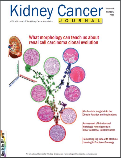
Into this milieu, Kapur and Brugarolas et al. have provided for the readership of Kidney Cancer Journal a thought-provoking review of their recent scholarship in assessment, analysis, and experimental modeling of the intratumoral heterogeneity of clear cell RCC (see Page 68 of this issue), touching on both molecular and histologic aspects13. Throughout, they draw an analogy between the well understood intratumoral molecular heterogeneity of clear cell RCC and the pathologist’s intuition that the tumor’s frequent intratumoral histomorphologic heterogeneity reflects this phenomenon. Recognizing that although clear cell RCC can be pathologically defined as a unique subtype, it is neither a single disease among different tumors, nor among clones within a single tumor. Drawing from their experience modeling the molecular heterogeneity of this disease and its progression14-16, the authors detail their recently reported comprehensive and quantitative histologic assessment of unique histologic patterns within (treatment naïve) clear cell RCCs13. These 33 patterns were sourced from three conceptual “axes”, specifically, architectural patterns, cytologic features, and histopathologic features of the tumor microenvironment. Data from detailed histologic feature assessment of over 500 clear cell RCCs were then correlated to key conventional pathologic parameters (grade, TNM stage, etc.), prognosis, and response to therapy. Remarkably, analyses based in their histomorphologic assessments recapitulated similar trends to those seen in prior studies based in genomic assessment.
Their project documents in unprecedented and quantitative detail the range of pattern heterogeneity between clear cell RCC tumors, with lower heterogeneity seen in smaller tumors than larger tumors, as well as a significant association between pattern heterogeneity and aggression. From the pathologist’s standpoint, intriguing, too, were observations of certain patterns predictive of survival, even after multivariable adjustment for the aforementioned, conventionally reported parameters. Striking to us, particularly, were the findings of analyses, based on adapting fundamental assumptions about tumor biology and evolution to the spatial relationships between histologic patterns. These analyses allowed them to infer even phylogenetic relationships between patterns. Thus they observed that the small nested or cystic/microcystic patterns, seen so routinely diagnostically, represent founder patterns, from which subclones outgrow with more aggressive patterns, converging on aggressive solid patterns. Preferential co-occurrence of certain patterns allowed inference that they were related, including observations with therapeutic relevance such as association of intratumoral lymphocytic infiltrates with sarcomatoid and rhabdoid features. Adroitly, they employed the paradigm of this disease’s natural history to assess inferences from their models, for example, validating patterns inferred to reflect aggression in primary tumors as present in later stages, such as thrombus formation.
Going forward, there are multiple implications of the authors’ ontological analyses of histologic patterns, and we suspect these go beyond their potential to nominate additional “univariate” prognostic parameters for pathologists to assess and report diagnostically. Awareness of the ontological relationships between different clones/patterns and establishing which reflect adverse prognosis or treatment resistance could be used to more capably manage RCC, not least by assisting in selecting the most appropriate tumor sample for precision medicine approaches. The power of (and accomplishments of) reductionist approaches like molecular studies to inform our understanding of the genesis of cancer and its progression should not be understated. Yet, we cannot help wonder as artificial intelligence/ machine learning approaches begin to assist in refinement, quantitation, and objectification of “next gen histologic” features like those assessed by Kapur et al., whether we are about to discover the prognostic and predictive power of higher order features of the cancer system, too.
REFERENCES
- Bennington JL, Beckwith JB. Tumors of the Kidney, Renal Pelvis, and Ureter. 2nd ed. Washington, D.C.: Armed Forces Institute of Pathology; 1975.
- Storkel S, Eble JN, Adlakha K, et al. Classification of renal cell carcinoma: Workgroup No. 1. Union Internationale Contre le Cancer (UICC) and the American Joint Committee on Cancer (AJCC). Cancer. 1997;80(5):987-989. https://www. ncbi.nlm.nih.gov/pubmed/9307203.
- Amin MB, Amin MB, Tamboli P, et al. Prognostic impact of histologic subtyping of adult renal epithelial neoplasms: an experience of 405 cases. Am J Surg Pathol. 2002;26(3):281-291. https:// www.ncbi.nlm.nih.gov/pubmed/11859199.
- Tickoo SK, DePeralta-Venturina MN, Harik LR, et al. Spectrum of epithelial neoplasms in end-stage renal disease: an experience from 66 tumor-bearing kidneys with emphasis on histologic patterns distinct from those in sporadic adult renal neoplasia. Am J Surg Pathol. 2006;30(2):141-153. doi:10.1097/01. pas.0000185382.80844.b1
- Aron M, Chang E, Herrera L, et al. Clear cell-papillary renal cell carcinoma of the kidney not associated with end-stage renal disease: clinicopathologic correlation with expanded immunophenotypic and molecular characterization of a large cohort with emphasis on relationship with renal ang. Am J Surg Pathol. 2015;39(7):873- 888. doi:10.1097/PAS.0000000000000446
- Magers MJ, Udager AM, Mehra R. MiT Family Translocation- Associated Renal Cell Carcinoma: A Contemporary Update With Emphasis on Morphologic, Immunophenotypic, and Molecular Mimics. Arch Pathol Lab Med. 2015;139(10):1224-1233. doi:10.5858/ arpa.2015-0196-RA
- Smith SC, Trpkov K, Chen YB, et al. Tubulocystic carcinoma of the kidney with poorly differentiated foci. Am J Surg Pathol. 2016;40(11):1457-1472. doi:10.1097/PAS.0000000000000719
- Williamson SR, Eble JN, Amin MB, et al. Succinate dehydrogenase-deficient renal cell carcinoma: detailed characterization of 11 tumors defining a unique subtype of renal cell carcinoma. Mod Pathol. 2015;28(1):80-94. doi:10.1038/modpathol.2014.86
- Hakimi AA, Tickoo SK, Jacobsen A, et al. TCEB1-mutated renal cell carcinoma: a distinct genomic and morphological subtype. Mod Pathol. 2015;28(6):845-853. doi:10.1038/modpathol.2015.6
- Kuroda N, Trpkov K, Gao Y, et al. ALK rearranged renal cell carcinoma (ALK-RCC): a multi-institutional study of twelve cases with identification of novel partner genes CLIP1, KIF5B and KIAA1217. Mod Pathol. May 2020. doi:10.1038/s41379-020-0578-0
- Motzer RJ. NCCN Clinical Practice Guidelines in Oncology: Kidney Cancer. 2018;Version 2. https://www.nccn. org/professionals/physician_gls/default. aspx.
- Turajlic S, Xu H, Litchfield K, et al. Deterministic Evolutionary Trajectories Influence Primary Tumor Growth: TRACERx Renal. Cell. 2018;173(3):595- 610.e11. doi:10.1016/j.cell.2018.03.043
- Kapur P, Christie A, Rajaram S, Brugarolas J. What morphology can teach us about renal cell carcinoma clonal evolution. Kidney Cancer J. 2020;in press.
- Wang SS, Gu YF, Wolff N, et al. Bap1 is essential for kidney function and cooperates with Vhl in renal tumorigenesis. Proc Natl Acad Sci U S A. 2014. doi:10.1073/pnas.1414789111
- Gu YF, Cohn S, Christie A, et al. Modeling renal cell carcinoma in mice: Bap1 and Pbrm1 inactivation drive tumor grade. Cancer Discov. 2017. doi:10.1158/2159-8290.CD-17-0292
- Brugarolas J. Molecular genetics of clear-cell renal cell carcinoma. J Clin Oncol. 2014. doi:10.1200/JCO.2012.45.2003


